
For any patient diagnosed with solid tumors, the day of surgery represents both a pivotal step toward survival and an agonizing test of emotional endurance. In cancer surgery, removing the visible tumor is only half the battle; the ultimate measure of clinical success lies in achieving “clear margins”—ensuring that no microscopic cancer cells are left behind along the resected tissue boundary.
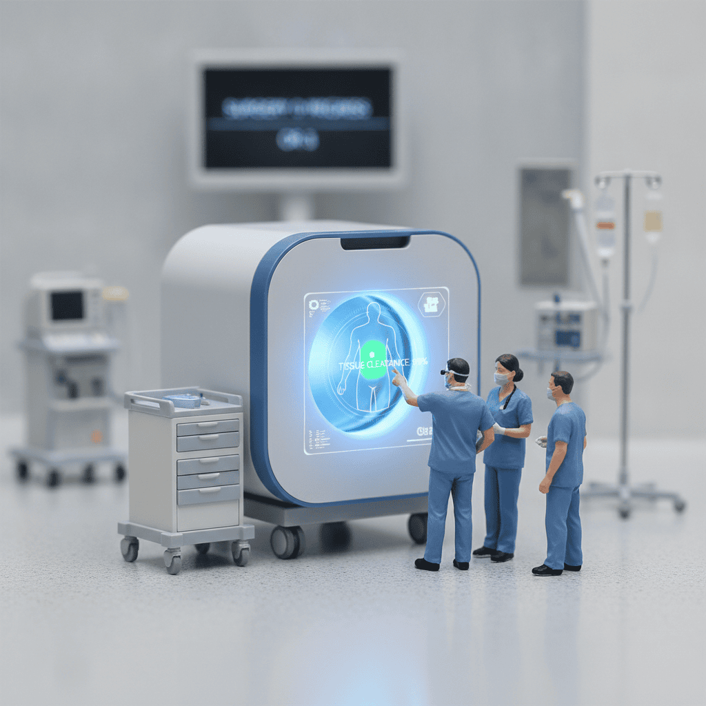
Traditionally, verifying margin clearance during surgery has been an agonizing race against the clock, often plagued by diagnostic uncertainty. Now, a groundbreaking technological breakthrough developed by South Korean biomedical engineers is poised to transform intraoperative surgical oncology: a ultra-compact 15-kilogram portable 3D Computed Tomography (CT) system capable of rendering high-resolution, volumetric margins in under 6 minutes.
The Silent Burden of Breast-Conserving and Oncologic Surgeries
Over the past three decades, oncologic surgery has steadily shifted toward tissue-sparing, organ-preserving approaches. In breast oncology, breast-conserving surgery (lumpectomy) has largely replaced radical mastectomy as the standard of care for early-stage malignancies, offering comparable long-term survival while preserving natural body contour.
However, preserving healthy tissue walks a razor-thin line with oncologic radicality:
- High Reoperation Rates: In the United States alone, approximately 300,000 breast-conserving procedures are performed annually, yet nearly 25 percent (around 75,000 patients) are forced to return to the operating room for a second re-excision due to positive or close microscopic margins.
- Psychological Trauma: Hearing that cancer cells were found on the margins days after an arduous operation inflicts severe emotional distress, compounding the fear of recurrence.
- Physical and Financial Toll: Reoperation delays essential adjuvant therapies—such as radiation and chemotherapy—while multiplying anesthesia risks, surgical complications, cosmetic deformities, and healthcare expenditures.
This systemic dilemma is not exclusive to breast cancer; head and neck resections, thyroid operations, and soft tissue sarcoma excisions face identical challenges where preserving critical nerves and structural anatomy must coexist with total tumor clearance.
The Critical Bottleneck of Conventional Intraoperative Assessment
Surgeons currently depend on two primary diagnostic pathways during surgery, each accompanied by substantial clinical limitations:
- Intraoperative Frozen Section Analysis:
- Although histologically informative, rapid freezing, slicing, and staining of fresh specimens takes between 45 and 60 minutes or longer per sample.
- Prolonged anesthesia escalates patient morbidity, while ice-crystal artifacts can distort cellular morphology, leading to diagnostic ambiguity in fatty or delicate glandular tissues.
- Conventional 2D Specimen Radiography:
- Operating room X-ray systems capture planar images within minutes, but they project complex 3D structures onto a flat 2D plane.
- Overlapping dense parenchymal tissue frequently obscures positive margins, leaving the surgeon unable to calculate the precise 3D distance between the lesion and the true resection boundary.
- Bulky Stationary Imaging Systems:
- Standard high-resolution specimen CT scanners weigh upwards of 400 kilograms.
- Their immense footprint, complex power requirements, and high acquisition costs make them impractical for integration within standard sterile surgical suites or standalone outpatient surgical facilities.
The Breakthrough: 15kg Point-of-Care 3D Specimen CT
Bridging the gap between speed, precision, and mobility, South Korean deep-tech medical startup Cobit—originating from KAIST (Korea Advanced Institute of Science and Technology)—has engineered an ultra-lightweight specimen CT scanner designed specifically for point-of-care intraoperative margin assessment.
Led by medical imaging specialists and former Harvard Medical School researchers, the team resolved the size-performance paradox through advanced detector design and proprietary computational algorithms:
- Unprecedented Speed:
- The scanner executes a complete 360-degree rotational specimen scan in just 2 minutes.
- Dedicated volumetric reconstruction engines assemble fully navigable 3D cross-sectional models in 3 minutes, delivering actionable diagnostic margins within a total of 5 to 6 minutes.
- Dramatic Miniaturization:
- At roughly 15 kilograms, the unit represents an astonishing 96 percent weight reduction compared to legacy 400-kilogram specimen CT units.
- It fits effortlessly onto standard surgical utility carts, allowing immediate point-of-care specimen loading without leaving the sterile theatre.
- Proprietary Color-Density Mapping:
- Unlike conventional grayscale radiologic outputs, the device applies intelligent color-mapping algorithms that visually distinguish subtle attenuation differences between fibrous stroma, healthy adipose tissue, microcalcifications, and malignant infiltrates.
- Surgeons can rotate the reconstructed virtual specimen in all anatomical axes, measuring millimeter-level clearance margins in real time.
Rigorous Validation and Expanding Clinical Horizons
Before moving into broad commercial adoption, medical imaging innovations must prove uncompromising sensitivity against gold-standard pathology:
- Preclinical Benchmarking: Rigorous trials utilizing tissue-mimicking phantoms confirmed high spatial resolution and volumetric accuracy within the sub-6-minute benchmark.
- Multi-Center Hospital PoCs: Collaborative clinical trials and proof-of-concept (PoC) studies are underway with leading Korean tertiary university hospitals, including Ajou University Hospital, Seoul National University College of Medicine Breast Cancer Center, and Chung-Ang University Hospital.
- Global Ambulatory Application: The compact architecture specifically targets Ambulatory Surgery Centers (ASCs)—rapidly expanding surgical facilities across North America and Europe where on-site pathology laboratories are scarce, yet same-day surgical discharge mandates immediate diagnostic certainty.
- Beyond Breast Oncology: Next-generation detector systems are already in development to expand chamber size, paving the way for intraoperative margin assessment in colorectal malignancies, pulmonary wedge resections, and complex endocrine neck surgeries.
The Future of Precision Oncologic Resection
The advent of rapid, intraoperative 3D specimen CT represents a paradigm shift from retrospective pathological confirmation to proactive, real-time surgical navigation. When surgeons can definitively confirm negative margins before closing the incision, the emotional nightmare of repeat resections can finally be eliminated.
As specialized surgical centers increasingly integrate high-definition imaging, minimally invasive robotics, and point-of-care analytics, cancer surgery enters an era where curative radicality and organ preservation no longer stand in conflict.
https://v.daum.net/v/20261030112917966?from=newsbot&botref=KN&botevent=a